


The Lipid Membrane: The Unsung Hero of Cellular Health
Authors:

Ed Kane
Introduction: Why Is the Cell Membrane Important for Cellular Function?
I found several pieces of my grandfather’s writing and I am so happy to share it with you - give you a peak as to why and how he started BodyBio.
At BodyBio, we believe health starts on the cellular level. Every one of your 40 trillion cells relies on a resilient, functional membrane—a lipid-based structure that defines cellular boundaries and orchestrates all intracellular activity. As my grandfather, Ed Kane, always reminded us, if you're good to your cells, your cells will be good to you.
- Jess Kane Berman
Inside the Lipid Bilayer: Why Is the Cell Membrane Important for Cellular Function
The cell membrane, also known as the lipid bilayer, is just 3 to 4.5 nanometers thick—but its influence is immeasurable. Formed by two layers of fatty acids, this barrier isn't merely passive—it’s the command center of the cell, embedded with proteins and ion channels that regulate everything from nutrient transport to immune signaling.
Imagine this: it would take 10,000 membranes stacked together to equal the thickness of a single sheet of paper. And yet, inside this ultrathin layer lies a dynamic, complex system critical to your survival.

Microtubules, Membranes, and Cellular Communication
The membrane doesn’t act alone. Within the cell, microtubules form a lattice that helps direct traffic, stabilize structures, and transmit energy and information. Thought leaders like Dr. Stuart Hameroff and physicist Sir Roger Penrose have suggested that microtubules may even play a role in consciousness through quantum wave activity.
While these ideas are still theoretical, the evidence points to a powerful synergy between the cell membrane and internal scaffolding—especially in highly specialized cells like neurons, cilia, and epithelial linings.
Essential Fatty Acids and Why the Cell Membrane Needs Them
Fatty acids are the raw materials of cellular membranes. Essential fatty acids (EFAs) like omega-3 (DHA and EPA) and omega-6 (LA and AA) are necessary for maintaining membrane fluidity and function. These nutrients cannot be synthesized by the body and must come from diet or supplementation.
When incorporated correctly, EFAs provide more than just structure—they regulate immune response, cellular signaling, and even mood. However, disruptions in fatty acid balance can cascade into a variety of degenerative disorders, including neurological decline and chronic inflammation.
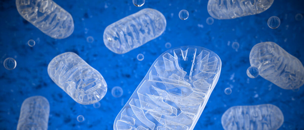
Phospholipids and Energy Production: The Role of Lipids in Mitochondria
Mitochondria are the energy powerhouses of your cells. But here's the secret: without a healthy lipid membrane, they cannot function. Nobel Prize-winner Peter Mitchell’s chemiosmosis theory revealed how the membrane enables the separation of hydrogen ions across mitochondrial layers to produce ATP—the energy currency of the body.
This membrane-dependent process is vital for life. And it only works when the lipid structure is intact and energized.
Cardiolipin and Mitochondrial Efficiency
Deep within the mitochondria lies cardiolipin, a rare and specialized phospholipid found exclusively in the inner mitochondrial membrane. Its unique structure—two phospholipid heads fused into one molecule with four fatty acid tails—allows it to play a critical role in stabilizing mitochondrial enzymes, maintaining membrane curvature, and supporting efficient energy production.
But cardiolipin’s function is only as good as its building blocks. And one of the most essential fatty acids required to construct healthy cardiolipin is linoleic acid (LA)—an omega-6 fatty acid that is often misunderstood but absolutely vital to mitochondrial performance.
Linoleic Acid: The Backbone of Functional Cardiolipin
In healthy cells, linoleic acid is the preferred fatty acid incorporated into cardiolipin. This specific configuration allows cardiolipin to optimally support mitochondrial function by:
-
Enhancing the structural integrity of the inner mitochondrial membrane
-
Facilitating the organization and efficiency of the electron transport chain
-
Supporting the binding of mitochondrial enzymes like cytochrome c
-
Promoting membrane fluidity and adaptability
When LA is deficient—or replaced with inappropriate, oxidized fats—cardiolipin becomes dysfunctional. The result? A cascade of mitochondrial impairment, reduced ATP production, increased oxidative stress, and cellular fatigue. This dysfunction is strongly implicated in a range of conditions, from neurodegeneration to metabolic disease and accelerated aging.
At BodyBio, we view unoxidized, clean linoleic acid not as a villain—but as a hero in the story of energy, healing, and cellular resilience. It is a cornerstone of our Balance Oil formulation and a critical nutrient for maintaining healthy mitochondrial membranes.
By prioritizing the right form of linoleic acid, we can help ensure that cardiolipin—and the mitochondria it supports—function at their highest potential.
Phosphatidylcholine and Aging: A Critical Link
Phosphatidylcholine (PC) is the most abundant phospholipid in the outer leaflet of the cell membrane—and one of the most important for cellular function, signaling, and integrity. With two fatty acid tails and a choline head group, PC forms the structural basis of the lipid bilayer, creating a fluid, flexible environment for membrane proteins and receptors to operate.
But this crucial molecule doesn’t stay static. In healthy, youthful tissue, PC integrates highly unsaturated fatty acids (HUFAs), particularly arachidonic acid (AA) and docosahexaenoic acid (DHA), at the Sn-2 position—making the membrane highly dynamic, responsive, and capable of rapid signal transmission.
As we age, however, levels of PC decline—replaced instead by stiffer, more rigid lipids like sphingomyelin (SM) and cholesterol. This shift decreases membrane fluidity, compromises nutrient transport, slows receptor signaling, and can impair organ and tissue performance across the entire body.
Aging and PC Loss: A Universal Phenomenon
In plasma membranes with low phospholipid turnover—such as the arterial walls and aorta—research shows a dramatic reduction in the PC/SM ratio. In aging tissue, this ratio may drop sixfold, and in advanced atherosclerosis lesions, SM can make up as much as 70–80% of total membrane phospholipids.
The result? A systemic loss of cellular elasticity and function, affecting every organ system:
-
Neuronal membranes lose their ability to communicate efficiently
-
Cardiac cells lose their rhythm
-
Immune cells falter in response
-
Skin and connective tissue become less resilient
-
Mitochondria, with their own PC-rich membranes, begin to fail
This isn’t just about structure—it’s about the functional life of every cell.
The Research: PC Rejuvenates Aging Cells
In one of the most powerful demonstrations of PC’s importance, researchers Yechiel and Barenholz at Hebrew University conducted a 20-day in vitro study using rat heart myocytes. These isolated heart cells were cultured in petri dishes and divided into three groups:
-
Group A: Received egg-derived PC starting on Day 6
-
Group B: Received no PC until Day 16
-
Group C: Received PC from Day 6–11, after which it was removed
The results were remarkable.
-
Group A maintained a consistent beating rate of ~160 beats per minute throughout the 20 days.
-
Group B experienced a steep decline by Day 12, some beating as low as 20 bpm—or not at all—until PC was reintroduced on Day 16.
-
Group C, which had PC removed mid-study, also saw a decline, mirroring Group B.
When PC was restored to Groups B and C on Day 16, both recovered fully within 24 hours—resuming their original rhythm and reversing the morphological signs of cell death.
This experiment showed, not just correlation, but direct causation: PC is a vital, non-negotiable component of cellular health and longevity.

PC in the Literature
A Medline search reveals over 37,000 peer-reviewed citations on phosphatidylcholine. Among the most compelling are:
-
Cui and Howeling (2002) – Demonstrated that perturbations in PC content lead to apoptosis (programmed cell death) and necrosis, and that restoring PC re-establishes cellular homeostasis
-
Yechiel and Barenholz (1985–1986) – Showed that membrane rigidity increases with age due to declining PC and increasing SM/cholesterol
-
Yehuda et al. (1993) – Suggested the optimal ratio of omega-6 to omega-3 (80:20) in membrane phospholipids for proper neuronal signaling and remodeling
These findings are not theoretical—they form the scientific foundation for BodyBio PC, our flagship phospholipid supplement. We formulate it to restore the lipid landscape of aging membranes, with a balance of high-energy essential fatty acids to revitalize every cell, from brain to heart to mitochondria.
The Membrane as the Brain of the Cell
Dr. Bruce Lipton, a cell biologist and pioneer in epigenetics, proposed that the membrane—not the nucleus—is the true control center of the cell. The DNA may house our genetic code, but the membrane is the processor and sensor, decoding signals and initiating activity.
A damaged membrane spells immediate death for a cell. Meanwhile, cells deprived of their DNA can continue living for weeks—proof of the membrane’s vital role in life.
Lipton calls it the “mem-brain”—and with good reason. Composed of over 70% fatty acids, this lipid-rich layer is the true intelligence of the cell.
Final Thoughts: Be Good to Your Cells
From the microcosmic elegance of the lipid bilayer to the life-sustaining role of mitochondrial membranes, the health of your cells starts and ends with fat—real, bioavailable, non-oxidized fat.
This is the legacy of Ed Kane, the founder of BodyBio. His vision was clear: to help people get better, one cell at a time. His work laid the foundation for everything we do today.
At BodyBio, we remain dedicated to his pioneering science, to exploring complex health challenges, and to formulating the highest quality supplements—like BodyBio PC and Balance Oil—that support your cell membranes, your mitochondria, and your mind.
Let’s be good to our cells. They’ll pay us back—a trillion times over.